The key to a healthy immune system is its remarkable ability to distinguish between the body’s own cells (self) and foreign cells (non-self). When the immune system identifies a non-self molecule it triggers an immune response. Anything that can trigger this immune response is called an antigen. An antigen can be a microbe such as a virus, or even a part of a microbe. Tissues or cells from another person also carry nonself markers and act as antigens.
In abnormal situations, the immune system can mistake self for nonself and launch an attack against the body’s own cells or tissues. The result is called an autoimmune disease. Some forms of arthritis and diabetes are autoimmune diseases. In other cases, the immune system responds to a seemingly harmless foreign substance such as ragweed pollen. The result is allergy, and this kind of antigen is called an allergen.
In abnormal situations, the immune system can mistake self for nonself and launch an attack against the body’s own cells or tissues. The result is called an autoimmune disease. Some forms of arthritis and diabetes are autoimmune diseases. In other cases, the immune system responds to a seemingly harmless foreign substance such as ragweed pollen. The result is allergy, and this kind of antigen is called an allergen.
|
|
|
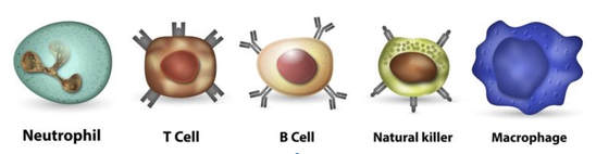
Cells of the Immune System
Leukocytes - White blood cells
Macrophages - These large cells "eat" invaders. Once ingested the microbe is broken down by enzymes within the macrophage. Macrophages can signal the body to flood the infected area with water which makes it easier for them to ingest invaders.
Neutrophils - release chemicals that kill bacteria. The chemicals are also dangerous to the neutrophils which are only able to live for a few days after releasing chemicals in the infected area.
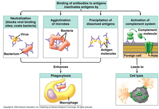
B cells - Also made in the bone marrow, B cells float around the blood looking for invaders. Each B cell can only recognize one type of antigen. When a B cell encounters an antigen that it recognizes it matures into a plasma cell and divides rapidly Plasma cells are factories for antibodies. They produces antibodies that are specific to the antigen the B cell originally recognized. Antibodies attach to antigens and signal the antigen for destruction. Antibodies can also surround an intruder, preventing it from attacking the host cells.
T cells - Made in the bone marrow, these cells travel to the lymph nodes where they are taught to recognize antigens. Antigens are parts of foreign cells such as proteins that are part of the foreign cell membrane. T cells are specific to a specific antigen. Thus tens of millions of different T cells are made in the hope of recognizing all the different types of antigens they might encounter. When a body cell is infected it presents antigens from the source of the infection on its surface. T cells don't recognize free floating antigens. Instead they recognize antigens that are presented on self cells. In this way T cells can kill infected self cells.
Natural Killer Cells - These cells kill foreign cells and infected self cells. Natural killer cells make a hole in the plasma membrane of the cell, water rushes into the pore and the cell bursts and dies. Natural killer cells kill anything that doesn't present "self proteins."
Macrophages - These large cells "eat" invaders. Once ingested the microbe is broken down by enzymes within the macrophage. Macrophages can signal the body to flood the infected area with water which makes it easier for them to ingest invaders.
Neutrophils - release chemicals that kill bacteria. The chemicals are also dangerous to the neutrophils which are only able to live for a few days after releasing chemicals in the infected area.
B cells - Also made in the bone marrow, B cells float around the blood looking for invaders. Each B cell can only recognize one type of antigen. When a B cell encounters an antigen that it recognizes it matures into a plasma cell and divides rapidly Plasma cells are factories for antibodies. They produces antibodies that are specific to the antigen the B cell originally recognized. Antibodies attach to antigens and signal the antigen for destruction. Antibodies can also surround an intruder, preventing it from attacking the host cells.
T cells - Made in the bone marrow, these cells travel to the lymph nodes where they are taught to recognize antigens. Antigens are parts of foreign cells such as proteins that are part of the foreign cell membrane. T cells are specific to a specific antigen. Thus tens of millions of different T cells are made in the hope of recognizing all the different types of antigens they might encounter. When a body cell is infected it presents antigens from the source of the infection on its surface. T cells don't recognize free floating antigens. Instead they recognize antigens that are presented on self cells. In this way T cells can kill infected self cells.
Natural Killer Cells - These cells kill foreign cells and infected self cells. Natural killer cells make a hole in the plasma membrane of the cell, water rushes into the pore and the cell bursts and dies. Natural killer cells kill anything that doesn't present "self proteins."
Antigens and Antibodies
|
Antibodies are proteins. As such, they are made of chains of amino acids that are folded into a specific shape based on the sequence and properties of the amino acids. Antibodies all have the same basic structure. Each antibody is made of four chains (shown in the image as blue and pink sections). There are two long heavy chains (colored blue) and two shorter light chains (pink/red). The specific binding site is found at the tips of the two arms, in a pocket formed between the light and heavy chain.
|
|
|
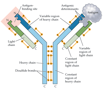
Figure 18.10, Purves's Life: The Science of Biology, 7th Edition (Found: http://www.hammiverse.com/lectures/43/2.html)
|
The binding site is composed of several loops in the protein chain that have very different lengths and arrangements of amino acids. Differences in these "hypervariable loops" form the many types of pockets in different antibodies, each of which bind specifically to a different antigen. The rest of the antibody is relatively uniform in structure, providing a constant recognition site for when antibodies interact with the rest of the immune system. In the image to the right you can see the variable region forms a specific shape that can bind with a specific antigen.
Antigens are molecules, such as proteins and carbohydrates, that can fit into the variable region of the antibody. This process is similar to how enzymes have a specific binding sites that match with specific substrates. |
ELISA Protocol
|
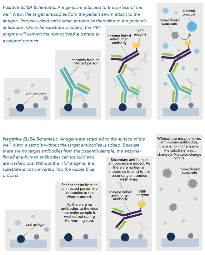
An ELISA (enzyme-linked immunosorbent assay) uses scientifically designed antibodies to detect if a particular substance, such as a viral antigen, hormone, or another specific antibody, is present in a sample.
The protocol involves binding an antigen or an antibody to a well plate. A sample is then added to the well plate. If the well plate was loaded with an antigen then the test will determine if an antibody for that antigen is present in the sample. If an antibody was loaded into the well plate then the test will determine if the antigen for that antibody is present in the sample. |
|
Adapted from Fred Hutch SEP guides: http://libguides.fredhutch.org/SEP/sep/elisa