|
A cell is the smallest unit of a living thing. A living thing, whether made of one cell (like bacteria) or many cells (like a human), is called an organism. Thus, cells are the basic building blocks of all organisms.
Several cells of one kind that interconnect with each other and perform a shared function form tissues, several tissues combine to form an organ (your stomach, heart, or brain), and several organs make up an organ system (such as the digestive system, circulatory system, or nervous system). Several systems that function together form an organism (like a human being). Here, we will examine the structure and function of cells. There are many types of cells, all grouped into one of two broad categories: prokaryotic and eukaryotic. For example, both animal and plant cells are classified as eukaryotic cells, whereas bacterial cells are classified as prokaryotic. Before discussing the criteria for determining whether a cell is prokaryotic or eukaryotic, let’s first examine how biologists study cells. |
|
Source: https://www.khanacademy.org/science/biology/structure-of-a-cell/introduction-to-cells/a/intro-to-cells
Microscopy
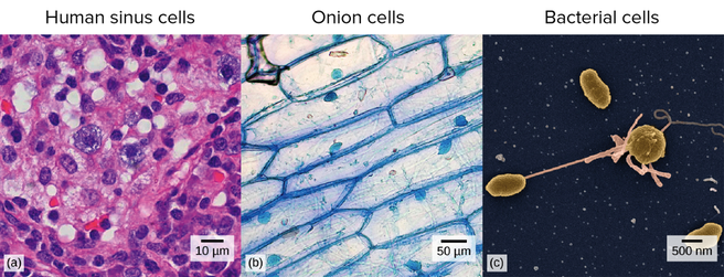
Cells vary in size. With few exceptions, individual cells cannot be seen with the naked eye, so scientists use microscopes (micro- = “small”; -scope = “to look at”) to study them. A microscope is an instrument that magnifies an object. Most photographs of cells are taken with a microscope, and these images can also be called micrographs.
Cells vary in size. With few exceptions, individual cells cannot be seen with the naked eye, so scientists use microscopes (micro- = “small”; -scope = “to look at”) to study them. A microscope is an instrument that magnifies an object. Most photographs of cells are taken with a microscope, and these images can also be called micrographs.
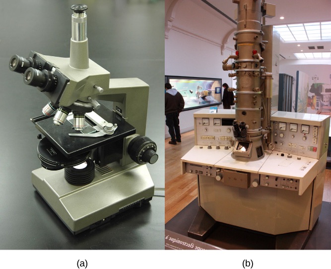
Figure 1 . (a) Most light microscopes used in a college biology lab can magnify cells up to approximately 400 times and have a resolution of about200 nanometers. (b) Electron microscopes provide a much higher magnification, 100, 000x, and a have resolution of 50 picometers. (credit a:modification of work by "GcG"/wikimedia commons; credit b: modification of work by evan )
Light microscopes
Most student microscopes are classified as light microscopes. Visible light passes and is bent through the lens system to enable the user to see the specimen. Light microscopes are advantageous for viewing living organisms, but since individual cells are generally transparent, their components are not distinguishable unless they are colored with special stains. Staining, however, usually kills the cells.
Electron microscopes
In contrast to light microscopes, electron microscopes use a beam of electrons instead of a beam of light. Not only does this allow for higher magnification and, thus, more detail , it also provides higher resolving power. The method used to prepare the specimen for viewing with an electron microscope kills the specimen. Living cells cannot be viewed with an electron microscope. As you might imagine, electron microscopes are significantly more bulky and expensive than light microscopes.
Cell size
At 0.1–5.0 µm in diameter, prokaryotic cells are significantly smaller than eukaryotic cells, which have diameters ranging from 10–100 µm. The small size of prokaryotes allows ions and organic molecules that enter them to quickly spread to other parts of the cell. Similarly, any wastes produced within a prokaryotic cell can quickly move out. However, larger eukaryotic cells have evolved different structural adaptations to enhance cellular transport. Indeed, the large size of these cells would not be possible without these adaptations. In general, cell size is limited because volume increases much more quickly than does cell surface area. As a cell becomes larger, it becomes more and more difficult for the cell to acquire sufficient materials to support the processes inside the cell, because the relative size of the surface area through which materials must be transported declines.
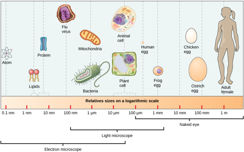
Figure 2. This figure shows relative sizes of microbes on a logarithmic scale (recall that each unit of increase in a logarithmic scale represents a 10-fold increase in the quantity being measured). Source: https://www.quora.com/Do-atoms-make-up-cells
All cells (Prokaryotes and Eukaryotes) share four common components:
Prokaryotes: Bacteria
- a cell membrane, an outer covering that separates the cell’s interior from its surrounding environment
- cytoplasm, consisting of a jelly-like cytosol within the cell in which other cellular components are found
- DNA, the genetic material of the cell
- Ribosomes, which synthesize proteins. However, prokaryotes differ from eukaryotic cells in several ways.
Prokaryotes: Bacteria
Source: https://6636.stem.org.uk/pathogens2.html
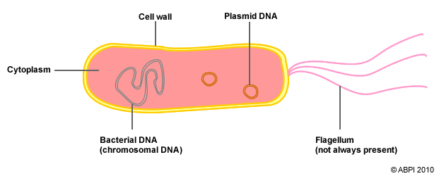
Bacteria have a cell wall that is different than plants. Rather than being made of cellulose it is made of peptidoglycan. This allows the wall to be more flexible. In addition, bacteria are different than eukaryotes because their DNA is not enclosed in a membrane bound organelle. There is no nucleus. Bacteria have two types of DNA, chromosomal (a long circular structure) and plasmid (small circular structures that contain non-essential DNA). Bacteria also often have a flagellum, a long, whip-like structure that helps them move.
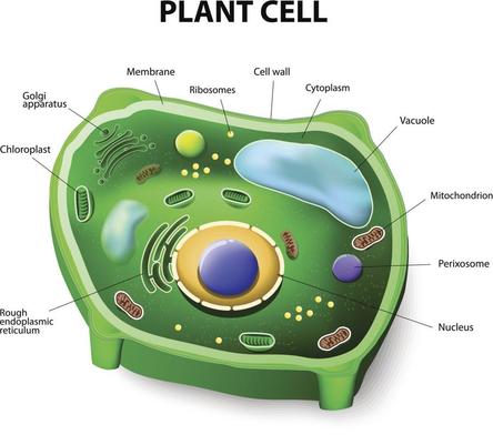
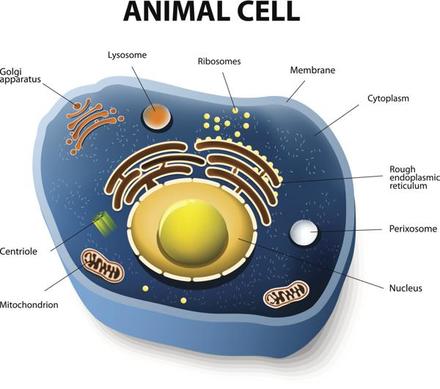
Eukaryotic Cells
Eukaryotic Cells
|
Source: https://biologywise.com/plant-cell-vs-animal-cell
|
Organelles
|
Cell Membrane
Cytoplasm Nucleus Mitochondria Cell Wall Chloroplasts Central Vacuole Plasmid DNA Flagellum Ribosomes |
Structure: Made of a double layer of phospholipids known as the phospholipid bilayer. A phospholipid is a lipid molecule composed of two fatty acid chains and a phosphate group.
Function: It is selectively permeable and controls the movement of substances into and out of the cell. The cell membrane is involved in facilitated diffusion, diffusion, osmosis and active transport. Structure: Made mostly of water, the cytoplasm is the liquid that fills the cell. Cytosol has a gel-like consistency that the organelles float in. A watery solution containing dissolved substances, enzymes and cell organelles. Function: The cytoplasm is the site of many chemical reactions and can act as a buffering solution ensuring appropriate conditions for enzyme function. Many metabolic reactions, including protein synthesis, take place in the cytoplasm. Structure: Surrounded by a membrane made of phospholipids, the nucleus contains the cells genetic material and nucleolus. Function: Controls and regulates cell activity. Often contains a dense spot of genetic material (DNA) known as the nucleolus where ribosomes are made. Structure: Surrounded by a double membrane, the inner layer contains cristae- folds that increase the internal surface area. Mitochondria contain their own DNA and ribosomes. Function: Performs cellular respiration. Structure: Semi-rigid structure outside the cell membrane made of cellulose in plants and peptidoglycan in bacteria. Cellulose and peptidoglycan are both carbohydrates. Function: Provides structure and support to the cell. The cell wall controls the volume of the cell by preventing too much water to be absorbed. Structure: Surrounded by a double membrane, the chloroplast contains a green pigment called chlorophyll, which is bound in a complicated series of proteins. Chloroplasts contain their own DNA and ribosomes. Function: Performs photosynthesis. Transforms energy from the sun into chemical energy in glucose. Only found in plants. Structure: Bound by a single membrane, the vacuole contains water and minerals for the plant. Function: The vacuole takes up most of the space in the cell. It regulates the amount of water in the rest of the cell. This is important for plants as they cannot control how much water they have access to. Only found in plants. Animal cells have small vacuoles not a large central vacuole. Structure: A plasmid is a small ring of circular DNA molecule often found within bacterial cells. Function: It is physically separated from a chromosomal DNA and can replicate independently. It contains additional genes that may or may not help the bacteria. Structure: Long hair-like structure protruding the cell membrane. Function: Assists the cell in movement. Found in Prokaryotes and some unicellular Eukaryotes. Found in sperm cells. Structure: Made mostly of protein and nucleic acids. There are to sub-units that fit together to make a ribosome. Function: Makes polypeptides by "reading" RNA and attaching amino acids to each other. After the polypeptide is properly folded it becomes a protein. Ribosomes make proteins. |
|
|
|
|
|
|
|
|
|
|
|
|