Redwood trees found in Yosemite National Park in California. Big? Of course. How do these trees grow so tall? It has a lot to do with a very efficient system to move water, sugars and other nutrients. But the first plants to have such a "vascular system" were not tall trees, but much smaller plants.
Vascular Plants
Vascular plants are known as tracheophytes, which literally means “tube plants.” The earliest vascular plants quickly came to dominate terrestrial ecosystems. Why were they so successful? It was mainly because of their tube-like vascular tissues.
Vascular Tissues
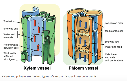
The vascular tissues for which these plants are named are specialized to transport fluid. They consist of long, narrow cells arranged end-to-end, forming tubes. There are two different types of vascular tissues, called xylem and phloem. Both are shown in Figure below.
Vascular Plants
Vascular plants are known as tracheophytes, which literally means “tube plants.” The earliest vascular plants quickly came to dominate terrestrial ecosystems. Why were they so successful? It was mainly because of their tube-like vascular tissues.
Vascular Tissues
The vascular tissues for which these plants are named are specialized to transport fluid. They consist of long, narrow cells arranged end-to-end, forming tubes. There are two different types of vascular tissues, called xylem and phloem. Both are shown in Figure below.
- Xylem is vascular tissue that transports water and dissolved minerals from roots to stems and leaves. This type of tissue consists of dead cells that lack end walls between adjacent cells. The side walls are thick and reinforced with lignin, which makes them stiff and water proof.
- Phloem is vascular tissue that transports food (sugar dissolved in water) from photosynthetic cells to other parts of the plant for growth or storage. This type of tissue consists of living cells that are separated by end walls with tiny perforations, or holes.
|
Credit: Mariana Ruiz Villarreal (LadyofHats) for CK-12 Foundation
Source: CK-12 Foundation License: CC BY-NC 3.0 |
This text was adapted from CK12.com. It is licensed under the Creative Commons (CC BY-NC 3.0). Original Source can be found here.
Leaf Structure and Function
Factories for Photosynthesis
A leaf is a highly organized factory – an organ constructed of several kinds of specialized tissues, each of which has its own duties. The product of the factory is no less than the food which supports nearly all life on Earth (although we must not forget that roughly half of Earth’s photosynthetic productivity is the province of algae and bluegreen bacteria, both evolutionary ancestors of plants.) In this section, we will explore how each tissue’s structure contributes to the production of food.
A leaf is a highly organized factory – an organ constructed of several kinds of specialized tissues, each of which has its own duties. The product of the factory is no less than the food which supports nearly all life on Earth (although we must not forget that roughly half of Earth’s photosynthetic productivity is the province of algae and bluegreen bacteria, both evolutionary ancestors of plants.) In this section, we will explore how each tissue’s structure contributes to the production of food.
|
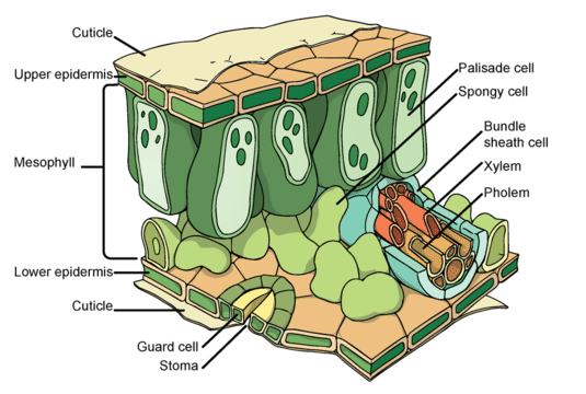
Credit: LadyoHats for CK-12
Source: http://commons.wikimedia.org/wiki/File:Leaf_anatomy_universal.svg License: CC-BY-NC-SA 3.0 |
The epidermis covers the upper and lower surfaces of the leaf. The epidermis mediates exchanges between the plant and its environment, limiting water loss, controlling gas exchange, transmitting sunlight for photosynthesis, and discouraging herbivores. The epidermis secretes a waxy cuticle, which restricts evaporation of water from the leaf tissue. This layer may be thicker in the upper epidermis compared to the lower, and in dry climates compared to wet ones.
The presence of the cuticle limits water loss, but also inhibits absorption of carbon dioxide and excretion of oxygen. These functions are served by stomata (singular, stoma), “little mouths” which regulate water loss, O2 release, and CO2 intake. In most leaves, stomata are more abundant in the lower epidermis, limiting water loss due to direct sunlight.
The presence of the cuticle limits water loss, but also inhibits absorption of carbon dioxide and excretion of oxygen. These functions are served by stomata (singular, stoma), “little mouths” which regulate water loss, O2 release, and CO2 intake. In most leaves, stomata are more abundant in the lower epidermis, limiting water loss due to direct sunlight.
|
The openings or pores in stomata are formed by two specialized guard cells. In daylight or high humidity, when CO2 is needed for photosynthesis and water loss can be minimized, the guard cells expand to open the pores. Closing the stomata is controlled by signals from roots when they sense water shortage.
|

|
An microphotograph (figure to the left) of a stoma shows the two guard cells which regulate its opening and closure to limit water loss, excrete oxygen, and absorb carbon dioxide.
|
Mesophyll (“middle leaf”) includes the tissues which build most of the interior of the leaf. These tissues conduct most of the photosynthesis for most plants, so most are made of thinner-walled cells with chloroplasts. In flowering plants and ferns, two different layers make up the mesophyll:
NOTE: Chloroplasts are structures inside of leaf cells where photosynthesis occurs. Chloroplasts contain chlorophyll, which is a green pigment that absorbs light energy. The chloroplasts are the green circles inside the palisades cells in the leaf diagram above.
The veins of leaves are made primarily of vascular tissue. An upper layer of xylem transports water and minerals from the roots and stem into and throughout the leaves. Recall that xylem is made of dead cells, with heavily thickened but pitted cell walls. The cells are arranged end-to-end, straw-like. Living cells of the lower layer of phloem, also arranged in bundles of straw-like columns but connected by sieve plates, transport sugars made in the leaves (“sugar sources”) to parts of the plants which need these fuels (“sugar sinks”). Like guard cells, vascular tissue harnesses the power of osmosis to accomplish movement without muscles. This feat is especially impressive because osmosis itself is a passive, entirely physical process.
This text was adapted from CK12.com. It is licensed under the Creative Commons (CC BY-NC 3.0). Original Source can be found here
.
- The upper, palisade layer captures most of the sunlight and carries out most of the photosynthesis. The columnar cells of the palisade layer contain many chloroplasts. Slight but precise separations between the cells maximize availability of the raw materials for photosynthesis by allowing diffusion of CO2 and capillary movement of H2O. Leaves exposed to high levels of sunlight contain as many as five layers of palisade cells, while shade leaves may contain only one.
- The lower spongy layer contains more rounded cells with fewer chloroplasts. The cells are loosely packed, separated by larger, airy spaces. This lower layer of cells is closely associated with the stomata, and the airy spaces allow diffusion of oxygen, water vapor, and carbon dioxide through the stomata when they are open.
NOTE: Chloroplasts are structures inside of leaf cells where photosynthesis occurs. Chloroplasts contain chlorophyll, which is a green pigment that absorbs light energy. The chloroplasts are the green circles inside the palisades cells in the leaf diagram above.
The veins of leaves are made primarily of vascular tissue. An upper layer of xylem transports water and minerals from the roots and stem into and throughout the leaves. Recall that xylem is made of dead cells, with heavily thickened but pitted cell walls. The cells are arranged end-to-end, straw-like. Living cells of the lower layer of phloem, also arranged in bundles of straw-like columns but connected by sieve plates, transport sugars made in the leaves (“sugar sources”) to parts of the plants which need these fuels (“sugar sinks”). Like guard cells, vascular tissue harnesses the power of osmosis to accomplish movement without muscles. This feat is especially impressive because osmosis itself is a passive, entirely physical process.
This text was adapted from CK12.com. It is licensed under the Creative Commons (CC BY-NC 3.0). Original Source can be found here
.